Bodi Empowerment
Back Pain //
MRI vs. CT Scan vs. X-Ray: Which is Right for You?

Deciding between an MRI, CT scan, or X-ray can be confusing, especially when you’re not sure which is the most appropriate for your specific health concern. This guide demystifies the process, helping you understand when each type of imaging is recommended and what to consider before undergoing a scan.
Understanding Your Options
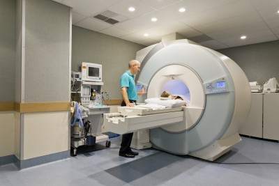
MRI (Magnetic Resonance Imaging):
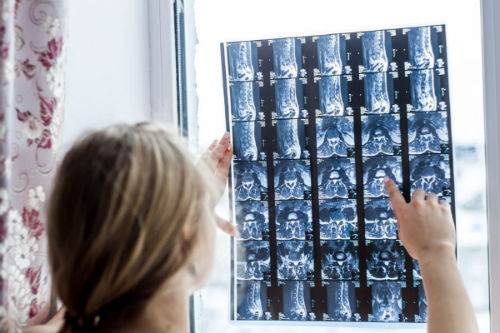
Utilizes magnets and radio waves to create detailed images of the body. Ideal for diagnosing issues related to soft tissues, discs, and the central nervous system, MRIs are more sensitive but less specific, revealing details that may not always correlate with symptoms.
CT Scan (Computed Tomography):
Combines multiple X-ray images to produce a comprehensive view of your body’s interior. Best for viewing bone injuries, diagnosing lung and chest problems, and detecting cancers. However, CT scans expose you to a higher dose of radiation compared to standard X-rays.
See Also: 4 Best Exercises For People With Sciatica
X-ray:
The quickest and most accessible form of imaging, perfect for examining fractures, infections, or abnormalities in the lungs. While X-rays are less detailed than MRIs and CT scans, they are invaluable for diagnosing specific conditions.
When to Choose Each Imaging Type
- MRI: Preferred for detailed images of soft tissues, spinal conditions, and joint problems. Essential for conditions like disc herniations, where soft tissue visualization is crucial.
- CT Scan: Optimal for quickly examining injuries or conditions that require a broader view of bone and organ systems. It’s particularly useful in emergencies.
- X-ray: Often the first step in diagnosing bone fractures or lung issues. It’s the go-to option for its speed and efficiency in revealing structural abnormalities.
Before Opting Imaging Tests, Ask Yourself Three Questions:
#1 Will the imaging pinpoint the cause of my pain?
The Paradox of MRIs and CT Scans in Diagnosing Pain
Have you ever wondered if an MRI or CT scan can pinpoint exactly where your pain originates? It’s a common question with a surprising answer: often, the answer is no. Here’s why understanding the capabilities and limitations of these powerful diagnostic tools is crucial.
The Limitations of Imaging
Misleading Results: Research shows that MRIs can reveal disc bulges and other spinal abnormalities in people without any pain—15% in 15-year-olds, 30% in 30-year-olds, and 60% in 60-year-olds. These findings challenge the assumption that visible issues on an MRI or CT scan always correlate with pain.
Sensitivity vs. Specificity: While MRIs are highly sensitive, detecting minute changes within the body, they are not always specific to the cause of pain. Dr. Christopher DiGiovanni notes that it’s rare to see a completely “normal” MRI, suggesting that abnormalities are common, even in asymptomatic individuals.
The Role of Pressure: Discs can change shape under different conditions, which means lying down during an MRI will not show the pressure that causes pain when sitting or standing. This discrepancy can lead to misinterpretations, where a patient’s symptoms don’t align with imaging results.
The Importance of Clinical Diagnosis
Beyond Imaging: A thorough history and physical examination are pivotal in making an accurate diagnosis. Sometimes, imaging findings may not reflect the patient’s experience of pain, emphasizing the need for a comprehensive evaluation by a healthcare provider.
Open MRI as a Solution: Open MRI machines, which allow for scanning in various positions, can sometimes offer a clearer picture of what happens to discs under different types of pressure. However, their availability and cost can be limiting factors.
Making Informed Decisions: MRI, CT, X-ray
Despite these challenges, MRIs and CT scans play a vital role in the diagnostic process, especially when differentiating between conditions like tumours and back pain. The key is to use them judiciously, informed by a thorough clinical assessment.
Remember, if you’re facing decisions about diagnostic imaging, discussing all aspects with your healthcare provider is crucial. Understanding the strengths and limitations of MRIs and CT scans can help you make informed choices about your healthcare journey.
#2 How will I know what is causing my pain then?
Unraveling the Mystery of Your Pain: Beyond Imaging
When you’re in pain, pinpointing the exact cause can feel like searching for a needle in a haystack. It’s natural to wonder if an MRI, CT scan, or X-ray can offer quick answers. However, the journey to understanding and addressing your pain often begins long before any images are taken.
The Central Role of Clinical Evaluation:
In-Depth Consultation: Your chiropractor or physiotherapist should start with a detailed conversation and physical examination, dedicating at least 15 to 30 minutes to understanding your symptoms, lifestyle, and any potential injury mechanisms. This initial step is crucial in forming a comprehensive view of your health status.
Diagnostic Expertise: Contrary to common belief, the true art of diagnosis lies not within the images produced by an MRI or X-ray but in the skilled interpretation of your history and physical examination findings. The overreliance on medical imaging has led to a decline in traditional diagnostic skills among some healthcare practitioners.
Formulating a Diagnosis: A proficient healthcare provider will consider your clinical presentation and may already have a primary diagnosis in mind, along with a few alternatives, based on your initial evaluation. The purpose of subsequent imaging tests like MRIs or X-rays is to confirm these suspicions rather than to discover them.
SEE ALSO: 6 Things You Should Look For In A Chiropractic Clinic: Chiropractic Clinic Best Practices
#3 Choosing the Right Diagnostic Imaging: MRI, CT Scan, or X-ray?

Deciding whether an MRI, CT scan, or X-ray is the best option for diagnosing your condition depends largely on your specific symptoms and the nature of your ailment. Understanding the strengths and applications of each imaging technique can guide you and your healthcare provider toward the most effective diagnostic approach.
When to Consider Each Imaging Option: MRI, CT Scan or X-ray
X-ray for Osteoarthritis:
- Ideal for Initial Diagnosis: X-rays are often sufficient for diagnosing osteoarthritis, characterized by morning stiffness that improves after movement.
- Cost-effective: Compared to MRI and CT scans, X-rays are more affordable and widely accessible for assessing joint wear and tear.
MRI for Disc Herniations:
- Superior Soft Tissue Imaging: MRIs excel in detailing soft tissue conditions, making them perfect for identifying disc herniations.
- Non-invasive Diagnosis: Before opting for an MRI, consider chiropractic care, extension exercises or Yoga Cobra, which can effectively treat many cases of disc herniation.
See Also: The Best Exercises For Your Herniated Disc
MRI for ACL Tears:
- High Sensitivity: MRIs provide detailed images of soft tissues, including ligaments, making them more sensitive for detecting ACL tears than physical examinations alone.
- Comprehensive Damage Assessment: An MRI can also reveal associated cartilage and ligament injuries, offering a complete picture of the knee’s condition.
CT Scan for Fractures Invisible on X-rays:
- Enhanced Bone Imaging: CT scans are superior for visualizing bone fractures that X-rays may miss, such as rib fractures.
- Critical for Complex Diagnoses: In cases where a detailed bone structure view is necessary, CT scans offer the clarity needed for accurate diagnosis.
Arthrograms for Cartilage Tears:
- Specialized Imaging: Injecting dye into a joint before taking an X-ray, MRI, or CT scan can highlight cartilage tears more clearly, essential for diagnosing meniscus, hip labral, and shoulder labral tears.
Key Considerations When Choosing Between An MRI, CT and X-ray
- Symptom-Specific Imaging: The choice between MRI, CT scan, and X-ray should be guided by the specific symptoms and the area of the body affected.
- Consult with Your Healthcare Provider: A thorough clinical evaluation is crucial in determining the most appropriate imaging technique for your condition.
- Cost vs. Benefit: Consider the cost and diagnostic value of each imaging option, especially when dealing with conditions that might be managed with less expensive alternatives.
Dangers of an MRI
An MRI machine has a powerful magnet that attracts most metallic objects. Therefore the danger lies with any implants that contain iron. Your implant can move and injure you physically or heat up and even burn you. The following are implants that are unsafe for MRIs.
- Older vascular stents
- Cochlear implants
- Brain-aneurysm clips
- Pacemakers or cardiac defibrillators
- Gastrointestinal cips
- Certain medication pumps (such as insulin pumps)
- Stainless steel spinal fusion screws and plates cause problems for the MRI and you. However, titanium screws and plates are fine. The stainless steel causes the digital picture to be degraded.
- Metallic objects like metal filings that have accidentally gone into the eye.
Gadolinium dyes are used in some MRIs, Gadolinium contrast dye is safe for most of you, but unsafe and can cause death in those with bad kidneys. If you are getting a procedure with Gadolinium your medical doctor should do a screening test for your kidney function.
Understanding the Radiation Risks: CT Scans, X-rays, and the Safety of MRIs
When it comes to medical imaging, understanding the risks and benefits of each option is crucial. While CT scans and X-rays are invaluable tools in diagnosing various conditions, it’s important to be aware of the radiation exposure involved. MRI scans, on the other hand, offer a radiation-free alternative for many diagnostic needs. Let’s dive deeper into the implications of choosing between these imaging techniques.
Radiation Exposure: A Comparative Look
Everyday Radiation vs. Medical Imaging
- On average, we’re exposed to about 2 millisieverts (mSv) of radiation annually from natural sources like rocks, the sun, and even aeroplane flights.
- Residents in high-altitude areas, such as Colorado, receive around 10 mSv due to less atmospheric protection.
CT Scans and X-rays: How Much Radiation Do You Get?
- A neck CT scan exposes you to about 6 mSv, roughly three years’ worth of natural radiation.
- A brain CT scan equals about 2 mSv, or one year’s worth of background radiation.
- A low back X-ray comes in at 1.5 mSv, about eight months’ worth of natural exposure.
- Neck X-rays and mammograms offer lower exposures, at 0.2 mSv and 0.8 mSv respectively, but these still add up over time.
Assessing the Need for Imaging
Before opting for a CT scan or X-ray, consider the following:
- Diagnosis Clarity: If your diagnosis is uncertain, imaging may be crucial.
- Treatment Impact: Will the scan results change your treatment approach? If not, reassess its necessity.
- Previous Scans: Remember, radiation exposure is cumulative. Evaluate the necessity of additional CT scans carefully.
- Cancer Risk: Those at high risk or with a family history of cancer should weigh the benefits and risks more carefully.
- Pain Localization: Can the scan accurately identify the source of your pain?
MRI: A Safer Alternative?
MRIs stand out as they do not use ionizing radiation, making them a safer choice in many scenarios. Despite advances in medical technology, it’s essential to consider the historical context—lessons learned from past practices, like the widespread use of X-rays in pregnant women during the 1960s, underscore the importance of caution and informed decision-making in medical imaging.
Making an Informed Choice: MRI, CAT scan and X-ray
Choosing between an MRI, CT scan, or X-ray involves a careful evaluation of your specific situation, guided by the expertise of your healthcare provider. By understanding the strengths and limitations of each imaging method, you can make informed decisions about your health care.
Tell us what you think in the comments below and like us on Facebook. This Toronto Downtown Chiropractor will answer all questions in the comments section.
Related Categories: Back Pain, Disc Herniation, Hip, Imaging, Low Back Pain, Shoulder, Surgery
Dr. Ken, has been recognized as the Best Toronto Chiropractor in 2024, 2023, and 2018, here in downtown Toronto. As a sports chiropractor, he excels in treating a wide range of conditions including concussions, temporomandibular joint disorders (TMJ), sports-related injuries, and spinal issues. Beyond his clinical skills, Dr. Ken is an accomplished athlete, having represented Ontario in the Canadian Judo Championships and completed the Toronto Marathon on two occasions. He employs the innovative C3 Program to provide targeted and effective care to his patients, ensuring a holistic approach to their well-being and athletic performance.


Hi there Dr. Ken. I had successful cervical disk fusion, 3 through 7 in 1998 due to an earlier whiplash. I have lived complication and pain free for fifteen years until getting rear ended again in July 2014. The doctors say both the CT scan and MRI reveal nothing that could be causing me such debilitating pain one year later. No nerve block test was done to rule out facet damage. They still can feel that is swollen, red, and hot to the touch, but insist there is no nerve or disk damage. I suffer daily with headaches, pain radiating down into shoulder blades and when I press on neck and shoulder it is very tender. If I suddenly, quickly move my neck, and even when I laugh, I will get sharp muscle spasms. My ear and jaw are also hurting and has locked on me several times. Often when I’m chewing, I can hear a crunching in my ear. How is it that I could be in such pain one year post whiplash if it was only a muscle sprain?? Is there no hope for me, I’m only sixty two and can’t imagine having to live with this level of pain. Thank you so much! Shelley
Author
Thanks for your question Shelley. I haven’t examined you but I would explore steroid injections and anti-inflammatories directed by your medical doctor or perhaps a different doctor. The redness, swelling and heat indicate inflammation. That’s very clear.
Hope that helps your neck pain.
Ii always feel unconfortable on my two legs just like some things run on and off on my blood.stream on both legs and its feels hot atimes when the weather is hot causing these these unconfortableness of putting on trousers feels like hell just like sometin is boiling and making my legs weak
Author
Thanks for your comment Folarin. Hope it gets better. It can be too many things. You really should be looked at by someone that can examine you. The comments section of this chiropractor in the MRI article is not the place for you.
Hope you get some help for your legs.
I had a whiplash-like injury while running on a treadmill about a year ago that left me with constant left-sided neck pain that travels to my jaw and head, dizziness, blurred vision and a distended left jugular. I only find comfort when lying flat on my back on a hard surface.
A cervical MRI showed minor disc bulges, which didn’t seem to concern my doctor. A brain MRI and MRA came back clear. A month of physical therapy improved some neck mobility but none of the pain.
Now my doctor has ordered a CT scan on the neck. Is there a better test? No one seems to know what they should be looking for. Despite desperately wanting to diagnose this pain I’d prefer some educated guesses before more testing.
Thank you for this website and all you do to help people!
Author
Thanks for your question Mary. It’s not a matter of a better test it’s a matter of finding a better therapist from my point of view. Go by reviews and referrals and find the best people in your area. Sound like the problem stems from the neck caused by nerve root irritation along with muscle and ligament damage. Also sounds like you have the jugular vein damage so it would be prudent to get a referral to a vascular surgeon. If there is vascular damage your vascular surgeon will probably send you for tests involving radioactive dye injected into your vein.
It’s not one thing causing your problem, it’s a constellation that were damaged in the accident.
Hope that helps your problems.
Thank you for your response and suggestions! The worst part of this process truly has been going to specialists, waiting months for an appointment only to have them say they can’t help. I’m very optimistic therapy will help given the proper regimen. I just worry about how long this has persisted with no improvement and what that means for my treatment.
Author
Thanks for the comment Mary. Sound like you are in the UK or Canada. While it’s better to get treated earlier it shouldn’t delay you too much. With any vein damage, if there is any damage to the vein I don’t know the prognosis.
Let’s put it this way. When people get in car accidents when they are significant enough it takes months and sometimes years to get better. You sound like you it hit your neck or head very hard.
You’ll have to be patient.
Hope that helps your whiplash injury. ( no more MRI or CT scans needed for now)
Hi Dr Nakamura,
I had a whiplash accident at work.
I was sent for a MRI by my spine specialist and will have my 1st physio this week after a month of resting at home and doing my own manual neck exercises at home.
C3-C4 mild disc osteophyte complex indenting the thecal sac. Severe right and mild left exit foraminal stenosis due to facet osteoarthritis compounded by uncovertebral hypertrophy is noted
C4-C5 disc osteophyte complex just abutting the cord. Moderate left and mild right exit foraminal stenosis due to uncovertebral hypertrophy and facet osteoarthritis is noted
C5-C6 there is disc osteophyte complex indenting the cord. Bilateral moderate exit foraminal stenosis due to uncovertebral hypertrophy is noted
C6-C7 Disc osteophyte complex indenting the thecal sac. Bilateral moderate exit foraminal stenosis due to uncovertebral hypertrophy is noted.
C7-T1 mild disc osteophyte complex indenting the thecal sac. The exit foraminal are patent
The spinal cord is normal in configuration and signal intensity
The paravertebral structures are unremarkable.
Conclusion
Multi-level degenerative disc disease is observed. This is worst at C5-C6 where the disc osteophyte indents the cord but there is no evidence of cord odema or myelomalacia
Author
Thanks for your comment Gejapathy. Your MRI report indicates that you have wear and tear or osteoarthritis that has taken decades to develop. Nothing visible on the MRI from the accident or whiplash.
Hope that helps your interpret your MRI.
Hi I’m April.. I have been dealing with pain in my lower back for many years. It gets worse when I do anything for a certain amount of time.like standing or sitting, laying to long. Usually within about 10 minutes and I have to adjust or do something else. I do have pain shoots down front of left leg only occasionally, numbness and tingling in toes. I am constantly adjusting. It’s frustrating. I’ve done physical therapy, epidurals with no improvement. I’ve had mri which my doctor says it just shows minimum degeneration. He says that a lot, like I’m making it up. It’s frustrating. Why would anybody pay that amount of money and making it up. I don’t know what to do. Any advice?
Author
Thanks for your question April. The last thing a doctor should rely upon is imaging like MRI, CT scans and X-rays. They have no correlation to pain by itself. That’s proven by research. I would consider a different doctor if he/she is basing his doubt on the MRI. (I don’t if that is true or not) You have to get someone that will do a thorough exam and history. For example minimal degeneration also coincides with disc bulges and disc protrusion. MRI aren’t that great yet. All they can tell is when they bulge, they miss the damage in the disc most of the time, in fact they catch disc damage approximately 12% of the time according to the literature.
When you sit and stand there is more pressure on the disc. You take and MRI when you lie down. The disc can bulge while you are sitting but disappear when you lie down and get your MRI. So if your doctor looks at the report and says you only have “minimal degeneration” than it seems your doctor is missing a lot.
Remember that is an example I gave you. I am not saying that is your diagnosis.
Hope that helps your understanding and how unreliable MRI and CAT scans and X-rays can be when solely relied upon.
Thank you so much for response. I do have disc protrusion, my Mri showed that also, but once again he says minimal. I appreciate your feedback. I may have to just find a new Dr. Thanks again.
Author
You are Welcome April
I had a car accident which caused me neck pain, almost immediately after, I went to the hospital the next day and had an x-ray, this showed there was something so they put a collar on me and sent me for a CT scan immediately, the CT scan showed “subtle impaction fracture in the superior endplate of C4”. About 6 weeks later I went for an MRI, the report indicates no impaction fracture. I have been in physio since the beginning and I’m taking Naproxen and Cyclobenzaprine, I am still very tender with limited range of motion, some pain from my shoulder blade down my right arm and tingling, this tends to happen when I’m sitting or driving usually. The C4 vertebrea is very tender to the touch, as my RMT found out recently, after lightly touching that area I suffered with pain for a few days. Which report would be more accurate with the impaction fracture statements the CT or MRI? Would treatment be different for an impaction fracture versus non impaction fracture?
Author
Thanks for your question Tammy. Keep in mind there are more modern and older versions of MRI and CT scans. As a general rule CT are better at “seeing” bone while MRI is better at “seeing” soft tissue especially discs. Both are very far from being perfect. For example when a surgeon looks at an MRI than after actually does a disc surgery the actual condition of the disc can be quite different. Only 12 % of annular tears are shown on MRI but are found during surgery.
So just because it didn’t show up on MRI doesn’t mean you healed. It means the MRI didn’t see it. I am sure your medical doctor or physio explained this to you, no? Often with an impaction fracture this causes the endplates (cartiladge) or the top of the vertebrae to break letting the contents of the disc to move into the vertebrae. This is sometimes called a vertical disc herniation as it mimics the symptoms of a regular disc herniation.
Try these exercises. https://www.bodiempowerment.com/cervical-disc-herniation-best-exercises-help-sore-neck/
These exercises must be done supervised or you will can get worse.
Hope that helps your impaction fracture.
I am feeling pain in my both legs during 3 months sometimes when I walk about 1and 2km i am feeling very fast pain in my both legs . when I wake up in morning I feels very huge pain
I had 2 spinal fusions at my lower back after my second operation my first ct-scan my doctor never saw that a screw has gone through my vertebrae i just had my second ct-scan i looked at my second disc and i can see the screw that went through my vertebrae i believe that is causing
my pain in my legs i go see my doctor in a few days
cat scan shows screw through my vertebrae how could a doctor missed this and not seen this
on the first c-scan
Author
Thanks for your question Jim. It’s not possible to tell if the screw is causing you pain without looking at the CT scan. Even then without examining you I wouldn’t be able to correlate the two. It’s pretty hard to miss a screw but that doesn’t mean it’s causing pain.
Instead of assigning blame I would concentrate on what you can do going forward. Unfortunately as I can’t tell what is causing the pain I can’t recommend any exercises. Why don’t you try the opinion of a neurologist recommended by your family doctor so you get an opinion that is unbiased. Recommendations from your surgeon will likely be someone he/she knows and will not want to contradict them.
Hope that helps your fusion surgery.
I stumbled across this forum looking for answers as I have so many times. I slipped on ice in early 2011 and have been in a significant amount of pain. Some days it brings me to tears and some days it is manageable. I have been to three physical therapists, a chiropractor regularly ( this provides temporary relief), an ortho, and family practitioners.
I always feel some degree of pain in my neck. This often leads to pain in my shoulders, upper back, and can cause intense headaches. I have had x-rays and an MRI (lying down). I have been told everything ranging from.. Nothing is wrong, degeneration in my cervical spine, and myofascial pain syndrome. Pain medication helps but due to having to work, it’s something I can only take at night and that is if I have it available as most physicians are reluctant to prescribe.
What step do I take next? Everything thus far (massage, pt, chiro) has helped a small amount, but I’m still in significant daily pain. Thank you!
Author
Thanks for your question Natasha.
Your diagnosis is not clear. However many cervical disc herniations can give you all those symptoms and is directly related to degenerative disc disease. You can try these exercises.
https://www.bodiempowerment.com/cervical-disc-herniation-best-exercises-help-sore-neck/
However there are no guarantees. It may or may not be your diagnosis. I am here to give you educational advice only. If you want a diagnosis you need to see another specialist as I cannot give you a diagnosis over the intenet.
If the pain gets worse with the exercises or increases symptoms further down the shoulder or upper back or into the headaches including, numbness, tinging, pins & needles or pain then you should stop and seek professional help nearby.
Hope that helps your possible cervical disc herniation.
Thank you. Would you recommend anything else as far imaging? Like I mentioned before, I have had xrays and an MRI. Is there another option that may provide better answers?
Author
Thanks for your question Natasha. MRI is the best that you can get. There are different setting off course like T2 which sees the water content in your disc. This is a normal procedure so most likely was done. Also the latest MRI might provide better resolution but you have to keep in mind even if gets a lot better you are not seeing what is true. MRI takes images while you are lying down. Most people with a disc problem have problems when sitting, standing or lifting at which time there is more pressure on the disc and thus more pinching of the nerve. Most MRI only provide images of you lying down. It’s doesn’t reflect what you do in everyday life.
I woudn’t get another MRI as it won’t provide any more information. A good health practitioner is more important.
Hope that helps your disc herniations.
Hi Dr.Ken,
Hoping you can help me.
I have a comminuted proximal humerus fracture to my right shoulder after falling heavily off my motorbike last August 20, 2014.
The second X-ray was taken on 3rd Nov reporting new bone growth evidence,but as yet a complete union of the fracture site..with fracture lucencies persisting.
The latest Radiographer reports of a Malunion?..with bony irregularity of greater tuberosity..but when I went to see the Orthopaedic Dr today he said after glancing at this X-ray..that the bone was healed fine and that I could return to work.
Whose report do I believe..?or should I get a second opinion..or perhaps an MRI or CT scan to confirm the true status of my fracture.
Thank you Dr. Ken
Author
Thanks for your question Anthony. Either the orthopedic surgeon didn’t look carefully at the X-ray or didn’t read the report. Malunion is serious. Go to another orthpedic surgeon to get surgery again. I would just move on. If the surgeon can’t see or can’t read it or simply is trying push you away as the surgery was unsuccessful we don’t know.
Many times the surgeon just did their best but the bone moved or surgery was very complex and the bones in many pieces in which case the arm often doesn’t heal.
Hope that helps.
Dr.Ken,
Reading your comments..I like to use holistic methods as much as possible.I am stuck.I am in constant pain in my lower back across the back area .around and below the hip line .I was given an antiinflammatory medicine but it did not help I have had an xray done nothing noted that’s causing pain .was thinking of going to a neurologist as my next step.I bought a heat patch for my back and that relieves the pain slightly.please any suggestions greatly appreciated.Thank you!
Author
Thanks for your question Cathy. Try these exercises first. https://www.bodiempowerment.com/4-exercises-lumbar-facet-irritation-lumbar-facet-syndrome/
If the exercises give you increased pain or start making your leg or buttock tingle or go numb then you should stop.
Hope that helps your lower back pain.
Hi. I have a line of pain all the way down the back of left leg. I have had sciatica before and this is different. I have delt with this for over a yr now. It is worse after standing or excercising or if I don’t get enough sleep. Pretty much constant other although some relief. Left knee also clicks a and hurts. About 6 wks ago I started having pain from groin down to left knee and pain in heel. Diagnosed as planter fasciitis. I have recently had hip xray, leg xray, leg mri. All were normal. I cannot go on like this. Dr gave cortisone injection in heel (which hasn’t helped much) and a night splint. Nothing for leg though. Any suggestions??? Thanks
Author
Thanks for your question Anna. It can be disc herniation or stenosis or spondylolisthesis. The diagnosis of plantar fasciitis is clearly wrong. You don’t get pain from the groin to the heel and call it plantar fasciitis. You clearly need a new doctor.
I recommend you seek a second opinion as exercises at this point may aggravate you.
Hope that helps your sciatica.
Hi, I have this sharp pain on my left side under my rib which radiates to lower back and numbness in abdomen and legs . Xrays and ultrasound are clear what could this be its really painful
Author
Thanks for your question Marilyn. You have a more complicated picture. I recommend you see your medical doctor as I cannot examine you or take a full history which will take a while.
I am a bodybuilder with an l5 fracture and herniated disc. I am awaiting a visit with a neurosurgeon for the first time on the 17th. Im not in constant pain but all it takes sometimes is getting out of a chair and I’m in pain for the rest of the week. It seems I then am crooked can’t straighten up after sitting and my pelvis and poster is hunched footwork ad to the right. I was told by my reg md that it is best is keep up the workouts to some degree in order to build my muscles for support. My question is can I still train? I need to be able to stress my muscles in order to the build and mold them. I can’t wear heels for long periods of time but I have a show coming up amd will need to. Will this end up hurting me worse? I am scared that I may be one squat away from hurting myself permanently. Is there anything I can do for building muscle in my lower back safely? Thanks!
Author
Thanks for your question Linda. You want your vertebrae that is fractured to heal properly before doing any exercises. If you had a fractured arm you can’t do light exercises to increase the muscle strength, until the bone is strong enough. Similarly you need to wait until the L5 fracture is healed enough.
Your competition should be secondary if you want to compete again in the future, in my opinion.
Hope that helps your L5 fracture and herniated disc.
Yes my 15 year old daughter was in an automobile accident a little over a month ago. She is having severe pain off and on all day on left side of neck and it radiates to her left top of shoulder. She has been on muscle relaxers and pain meds for a little over a month now with no relief. They did xray ct scan and MRI but nothing shows up. Its to the point she can’t hardly sleep at night and has already missed 11 days of school. I can feel the swelling on the left side of her neck. What should we do. It is frustrating watching her go through this pain after all the medication physical therapy and no relief.
Author
Thanks for your question Tim. Sounds like your daughter is in a lot of pain and that you care for her dearly, like most parents. When you get in a car accident it damages the ligaments, tendons, disc and muscles in the neck and sometimes even the bones. Sounds like the bones weren’t affected otherwise they would show up on the MRI. The swelling can be a muscle spasm or actual inflammation that is still on-going. If it is inflammation you should keep putting ice on it especially if it is red and swollen. The more likely scenario is that there is a muscle spasm from the damage done to the muscle possibly due to a tear. (A lot of things don’t show up on X-rays).
Also there is the possibility of a disc protrusion or herniation. The reason is the MRI are done while laying down, which puts the least amount of pressure on the disc. Well life involves sitting and usually bending forward when we are looking at the compute screen. There is simply more pressure when you do these things and the disc will stick out more. The MRI simply won’t see it because she isn’t sitting while doing an MRI or sitting with her neck bent forward.
The first thing I would do is get another opinion and go to a doctor that doesn’t depend only on MRI etc.. first make a clinical diagnosis then use the MRI and X-rays etc.. that is already there to confirm things.
If you get the right diagnosis it is easier to treat and get results. A disc herniation treatment is different from a muscle spassm treatment which is also different from a tear.
Hope that helps your daughter’s neck pain.
Hello,
My mother has an exposed nerve in her lower back close to the spine. It’s causing her severe pain all the time. She has been going to a specialist where they have been giving her aseries of needles. They now say that some spinal fluid is leeking. One Dr. Said surgery is not needed. Another said she may have too.????
Author
Thanks for your comment Bruce. Do you mean she has an open wound when you say she has an “exposed nerve” causing the spinal fluid to leek. If she does it’s most important to prevent infection first. Exercises are secondary. This is outside my scope so I have not examined her so it’s not possible to say if she needs surgery or not.
Hope your mother recovers soon.
Dear Dr Ken
I am reading your comments about back pain. I have been having pain during the night only since last December in my right leg mostly from my top right limb to my ankle and going up to my knees. Lately I have become worse and had paid during the day too and I stopped practically all activity. Now I went to a chiropractor and he did some stretches etc. He thinks that it is L2 and L3 problem when he saw the X ray. I am better and now my pain during the night is less severe. Now the paid moved upwards just behind my knee and discomfort on my buttocks. I am doing exercises putting my legs up right and left and when sitting down putting my back arch more to the front. Should I do more exercises that you suggested above like the cat? In the morning sometimes I am bad but then release by time. It is worse during the night and I could not sleep properly.
Thanks for your concern.
Regards
Lucy
Author
Thanks for your comments Lucy. Since the chiropractic treatments and exercises are currently working I think you should stick with that person. If they stop working or you plateau for a while you can do the exercises outlined here. https://www.bodiempowerment.com/sciatica-4-best-exercises/ Providing the diagnosis is correct
I am not sure the you have sciatica related to the lower back from the description of your pain. If is it related to sciatica from the lower back and it is a disc that is going backwards than the exercises in link above are appropriate.
Hope that helps your leg pain / sciatica.
Hi, I am Autumn. My husband has had back surgery for a crushed disc at the L5S1 area. He is almost 8 months post surgery and still has severe pain down both legs making it difficult to walk or stand for long periods. He has pain and tingling and loss of balance. He is still have spasms up and down his back. The Dr has done both MRI and CT and said that they were normal. What do you think we migt need to do for him. The Dr said this is as good as he will get…
Author
Thanks for your question Autumn. Surgery should usually help but if it didn’t then that is a very bad sign. Having said that there may be more minor goals that are attainable but definitely not guaranteed.
Try doing these exercises except for the sphinx and cobra and standing extensions. If he gets sharp pain, more pain while doing the exercises or pain for more than 10 minutes after doing the exercises than the exercises are not for him.
https://www.bodiempowerment.com/herniated-disc-part-2-the-best-exercises-for-your-herniated-disc/
The goal in his case would be to get him to stand 10 minutes longer or sit a little longer before the pain gets worse.
Also get a second opinion from a neurologist not a neurosurgeon. This way there is no conflict of interest as neurologists cannot do surgery so can only recommend someone else.
Hope that helps your husband’s disc.
I have pain radiating down my leg into my foot. I was diagnose with a large os trigonum in my right ankle yet the doctor said it was a normal right foot. A Os trigonum is a small round bone posterior to the talus. So how can a large Os triginum be normal? I have sclerosis bilaterally in my sacroiliac joint. What should be the diagnosis?
Author
Thanks for your comment James. Your concern is that the since the Os Trigonum is large, to you it doesn’t sound like the foot is normal even though your doctor says its normal and the X-ray report likely says it is normal.
The problem when the OS Trigonum is large is that it can get pinched between your ankle and heel bones. Usually the pain is triggered by walking, jumping and pointing your toes down. You can try treating it by doing trigger point work on the leg (calf).
Trigger points is simply pushing on a muscle with your thumb and finger on a sore spot on your muscle for about 10 seconds. So you simply push hard on calf muscle where it sore from your achilles all the way up to the knee joint. Work your way up in rows until you treat the whole muscle. When you come across a particularly painful and sore spot push for a longer period of time until it starts to feel better.
Warning: you shouldn’t do this if you have any of the following like Diabetes, previous high cholesterol, previous stroke, heart attack, high blood pressure and smoke a lot. The reason is you can dislodge a clot if you are susceptible to clots (those that have the above symptoms are more likely to have leg clots). Leg clots can be dislodged and cause a stroke.
If your pain actually feels likes it comes from higher up in your leg and radiates down than you need to get your lower back examined.
Hope that helps.
Hello I have been having extreme pain in my shoulder neck and head areas. Only ever in the right side. I have constent pain along with pain spikes that cause me to blackout due to the pain. I have had no luck getting any help at all. I am wondering if a mri or other scan will help show any possible problems. And what scan would be best for the upper back into the head. To show the most details and everything to hopefuly find out the cause to this long term extreme pain problem. I have the current option for a mri even tho I can barely afford it. So I want to find out if this will help pin point a possable cause to the extreme pains. Or be yet another great waste of money and time. Any help or thoughts at all would be great. If possible please reply via email. Thank you
Author
Thanks for your comment David. Can you explain where your pains are in your shoulder neck and head.
For example the pain might start on the right side of your neck and spread to your shoulder and upper arm and the back of your head or your pain might start at the top of your head spread down to your neck and eventually your upper back near your shoulder blade. What makes it worse, better? Does it the pain go radiate into the shoulder and into the hand? Give me more details.
My job is not to diagnose you but give you a possible idea as to which machine can possibly help you.
Waiting for your reply.
Thank you so much for your reply and help. The pains started about 5 years ago acting like a migrain with extreme pain in the right side of the head like a extreme migrain. And over the years has spread to include my whole right side from botom of shoulder blade threw the head. I am in constent pain in my neck and shoulder right side only. That pain is best described as a ice pick attached to a clamp squezzing down on certain points. In the upper shoulder area or any section of the spine from that point up threw the atlas, axis, and skull opening for the spine. Nothing seems to trigger the pain it jus comes and gose as it pleases. And very little will help ease the pain. Now this pain dose get way wors genraly when the pain spikes happen its in the upper shoulder, neck area. Or the base of my skull around the atlas, axis, base of skull. This pain starts out as a similar pain as stated earlyr. But quickly gets way wors filling like the most undescribale pain I could ever imagen. Which quickly spreads from which ever starting point threw to include the whole right side only. From mid, lower back up threw the entire head. The pains will cause a simulated but much wors migrain pain in the head which causes right eyes light sinsativatie. Excess tear production, excess mucus production which tends to clog my sinius adding sinius pains. I have to contiuly try to force open my eye to release the tear build up aswell as force clear my sinius to relieve that pressure pain. Whenever I can get a basic control of a pain spike or get my sinius cleared in the process. The pains grow more extreme and undescriable until a point I blackout from it. When I blackout I go from unconfortably moven around in pain trying to find a postion to relieve it. To waken up 2-4 hours later genraly startled awake by a noise or sumthing touching me like a pet. I do not lose control over bladder or bowels when blacked out. I seem to simply blackout from the pains to wake up few short hours later. When I do come to again I genraly either fill extremely tired and am able to go to sleep for a few hours. Sometimes waking up in pain again sometimes not. Or sometimes ill wake up filling somewhat rested and ready to go again with only genral disconfort or pain. All the disconfort and extreme undescribale pains are only ever in my right side almost perfectly down the center of my spine. And there has only ben 2 times which the pains crossed that line to include my left side. Both times it started out the same as described above but quickly taraveld across my upper back and neck to include the left side. When that happened both times it spread up into my head to include a extreme migrain filling. Aswell it traveled down my back and arms to make them fill extreme pains mixed with numbness and tingling. And would cause my hands to lock up in pain aswell balling up in a weird manner, not a full balled up fist as sum fingers did not cruel up. Those 2 pain sessions where by far the wors and scarest but only lasted 30-45 mins and happend 3 years apart the last one being about a year ago. The other described undescriable pain sessions last anywhere from 30mins to a full day or more. The wors being 4 days unable to sleep more than a hour due to the pains and no more than a half hour at a time where the pains where not near blackout levels with 5 blackouts in the 4 day stretch and that was about a year n half ago. I am afraid I can’t give you much true medical answers as I have not recived any attion or help as much as I have tryed to get it. And have had to start reserching and trying to figure things out on my own. When I found your very helpful site and desided its worth a shot to ask for sum knowlage n help. I am looking foward to your next reply and help, knowlage. And will contiue to give you all the info I can that will better help you out in helping me a little. Thank you so much for your help kind sir.
Author
Your pain could be related to your neck or your head. Really your pains are too complex without an examination. You should seek out a professional health care worker. I would consider a neurologist due to the nature of your pain. After five years it is possible that it is a neck problem that has centralized. Meaning that having so much pain for so long changes your nervous system so that very little provocation gives you extreme pain.
Please seek out proper professional care in your area as your case requires hands on examination. I wish you the best of luck.
Well honestly the only reason this has gone on this long. Is due to not being able to get proper attion and care all this time. The only thing I can do is keep trying, armed with this new helpful knowlage you have provided me. Thank you so much for you fast respones, intrest, knowlage, and careing. You are a good man keep up the good work.
Thanks Dr. Ken for introducing such an effective method of self treatment for us and hope that you will bring more effective way for us in near future as well. So will look forward once again to see your next update and hopefully that will be more excitement. Really appreciate able!
Author
You are welcome William. Since you are commenting about the MRI article, I want to remind you to make sure you actually need an X-ray or MRI. Many doctors including chiropractors feel pressured to give X-rays and MRI when either the patient demands it, when a patient isn’t getting better (not a bad reason), or they want to pay for the MRI or X-ray machine that they bought. An X-ray machine can now run $100 000 -$200 000 while an MRI can cost $2 million dollars. Just make sure they are following the criteria outlined in the article.
hi doc is it true that some doctors and ragiologist are not able to read xray reports correctly
Author
Thanks for the comment Seth. General practitioners and family doctors are trained during medical school to read x-rays, CT and MRI scans. During residency (internships) there is usually further education. However at least in Canada most family doctors come to rely on the reports made by radiologists and don’t even look at X-rays. Those doctors that also work in hospitals may be an exception. My guess is, that the US is similar. For Europe I think it varies according to country.
So if a family doctor doesn’t read a X-ray or MRI for 10 years they are not very good at it. Radiologist on the other hand read X-rays and MRIs everyday so are generally good at it.
Even with experienced radiologist you can get X-rays, MRI reports that have missed, sometimes obvious problems. There probably many reasons for this ranging from competency, boredom, senility for the soon to be retired radiologists. In other words they are human and make errors sometimes. To avoid this you can always have an MRI or CT Scan or X-ray re-read by another radiologist for a second opinion or a chiropractic radiology specialist with the designation “DACBAR”.
Hope That Helps.
Dr. Ken hello how’s it going My name is Rodolfo i am 20 years old and i have been having pain on my lower mastoid bone and goes down the side of my neck for about 2 years now, i had an ultrasound performed on that side of my neck and the doctor could not find anything, said everything looked normal. The pain persisted so about a week ago i had an xray of my mastoid performed and the doctor told me everything looked normal again but still the pain persists. I am going crazy with this pain , i notice the pain kind of subsides when i lay down or stretch my neck. Pain increases when im standing. The doctor has ordered a ct scan but i dont really want to expose myself to so much radiation. My main concern is tumors and cancer . Im so scared sir, but my doctor insists its most likely due to a bad muscle on the side of my neck. I don’t know what to do anymore i really dont want a ct scan i rather get an MRI but im not sure if it will show anything. What is your best advice doctor i am so scared i am only 20 years old and i can’t live my normal life. I stopped the gym, i can’t concentrate in school, i can’t work normally without thinking of the pain. I feel horrible please give me some advice.
Author
Thanks for your question Rodolfo. First as you know by now ultrasounds and X-rays, MRI and CAT scans even when they find things it often doesn’t correlate with the pain. You need to go to a good chiropractor or good physiotherapist or an osteopath or even a massage therapist. They are trained to look at muscles, bones, nerves, ligaments and schooled in how to treat those conditions. A medical doctor is trained to diagnose those things but not treat them. They can recommend medication or refer and unless they are a specialist. A specialist like a physiatrist is a medical doctor trained to treat these conditions. Orthopedic surgeons generally do not treat manually, usually sticking to medication, injections or surgery.
The general rule is that you use conservative treatment like physiotherapy, chiropractic or osteopaths first. If that doesn’t work use injections and medications.
You haven’t tried conservative treatment unless you left that part out. That’s an important first step to diagnosing and getting things better.
Hope that helps your neck (mastoid area) pain. Keep in mind the sternocleidomastoid and longissumus capitus muscles attach to the mastoid. Usually you treat the muscle the pain goes away. With person like yourself it will likely take longer as you are focussed on the pain and thinking the worst. Most of the times it isn’t the worst. What you are doing is called catastrophizing. This only makes any problem worse sometimes multiple times worse than the actual problem.
Find yourself a good chiropractor or physiotherapist, get them to calm you down then treat your problem.